Posted on — Leave a comment

Bacteriostatic Water for Peptides: Lab Handling Guide

Bacteriostatic water for peptides is one of the most searched lab-handling topics, and for good reason: most research peptides ship as a lyophilized (freeze-dried) powder, and the diluent you choose affects how the material behaves once it is in solution. This guide explains what bacteriostatic water is, how it differs from sterile water, and the handling, labeling and storage practices laboratories use to keep reconstituted research peptides consistent from the first draw to the last.
What Is Bacteriostatic Water for Peptides?

Bacteriostatic water (often shortened to BAC water) is sterile water that contains 0.9% benzyl alcohol. The benzyl alcohol acts as a preservative: it does not sterilize a solution, but it inhibits the growth of most bacteria that could be introduced when a vial stopper is punctured more than once. That is why BAC water is the standard diluent in many labs that need to draw from the same reconstituted vial several times.

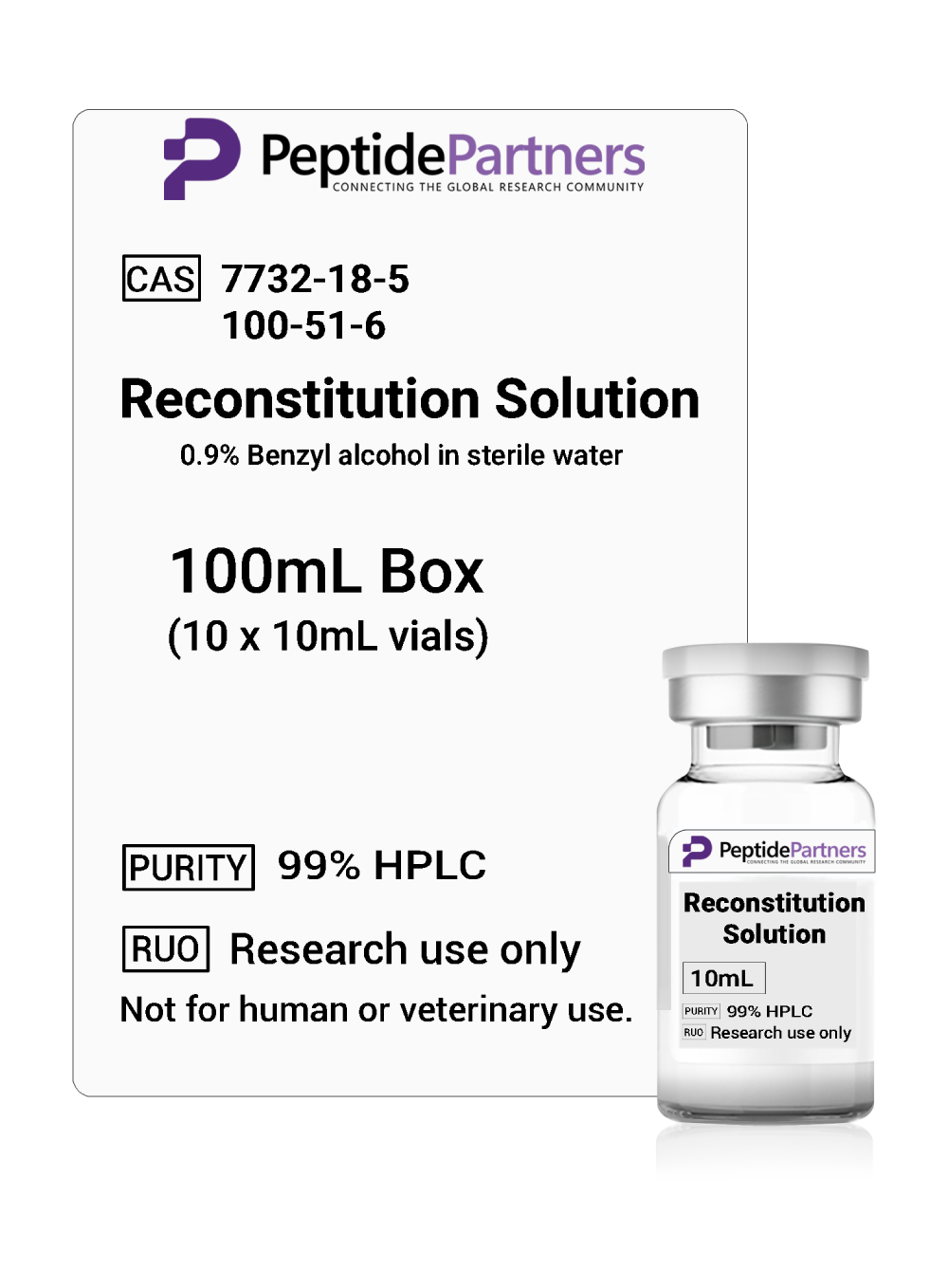
Our Reconstitution Solution (BAC water, 10mL vials) is supplied for exactly this purpose: dissolving lyophilized research peptides for laboratory work.

Bacteriostatic Water vs Sterile Water

Both diluents start from sterile, pyrogen-free water, but they are suited to different workflows:

  • Bacteriostatic water: contains 0.9% benzyl alcohol, designed for multi-draw vials, and is the common choice when a reconstituted peptide will be sampled repeatedly over days or weeks.
  • Sterile water: contains no preservative, is intended for single use, and is typically chosen when a protocol requires a preservative-free solution or the entire vial will be used at once.
  • Other buffers: some peptides are poorly soluble in water alone, and published methods may call for dilute acetic acid or a specific buffer. Always check the literature for the compound you are studying.

For most routine multi-use research workflows, bacteriostatic water is the practical default because it adds a layer of protection against contamination between draws.

Why Lyophilized Peptides Need Careful Reconstitution

Lyophilization removes water from a peptide so it remains stable during shipping and storage. Once the powder is returned to solution, the peptide becomes more vulnerable to degradation from heat, light, agitation and microbial contamination. Gentle, consistent handling helps preserve the integrity of the sequence you paid to have verified.

Good laboratory practice for reconstitution generally includes:

  • Allowing a refrigerated vial to reach room temperature before opening to limit condensation.
  • Wiping vial stoppers with an alcohol swab and using sterile equipment for every puncture.
  • Introducing the diluent slowly down the inside wall of the vial rather than directly onto the powder.
  • Swirling or rolling gently until dissolved, and avoiding vigorous shaking, which can cause foaming and aggregation.
  • Visually inspecting the solution: it should be clear, without particulates or cloudiness.

Calculating Concentration for Lab Records

Concentration is simply peptide mass divided by diluent volume. For example, a 10mg lyophilized vial reconstituted with 2mL of bacteriostatic water yields a 5mg/mL solution; the same vial with 1mL yields 10mg/mL. Choosing a volume that produces a round number makes downstream calculations in your experimental protocol easier and reduces transcription errors.

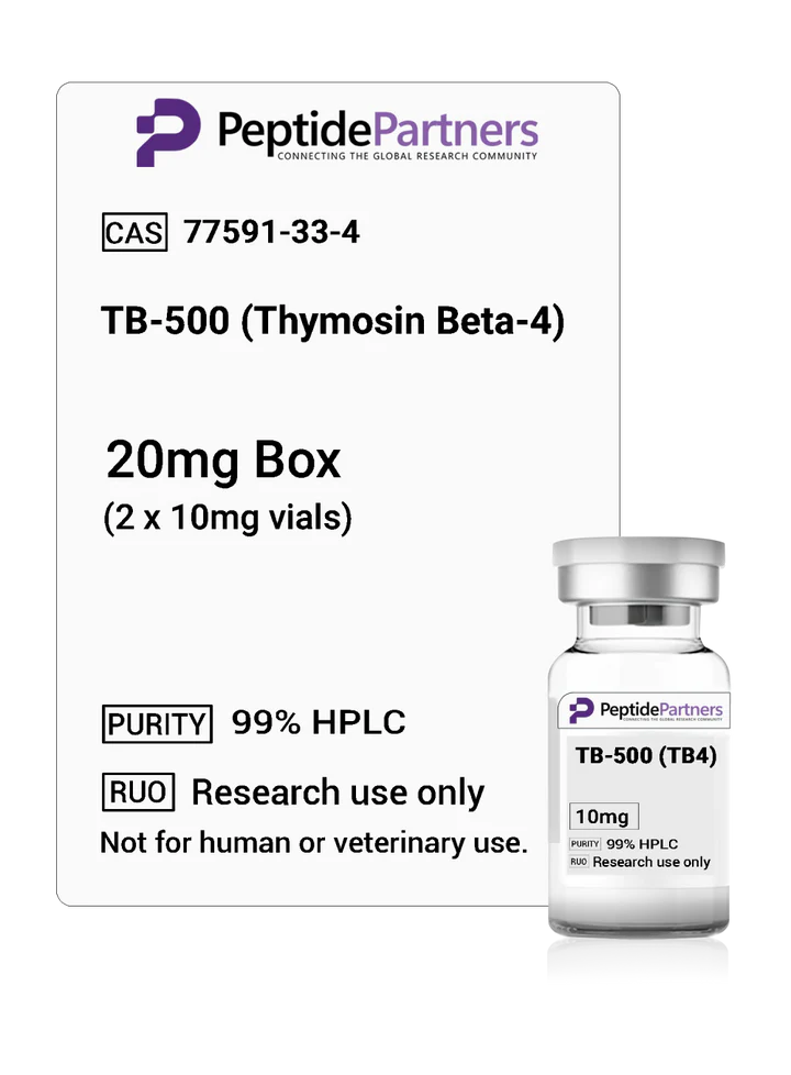
Record the compound, lot number, diluent, volume added, resulting concentration and reconstitution date directly on the vial label and in your lab notebook. Our 10mg research vials, including TB-500 (TB4) 10mg vials, Sermorelin 10mg vials and Thymosin Alpha-1 10mg vials, all make this math straightforward.
Storing Reconstituted Research Peptides

Storage conditions are one of the biggest variables in peptide stability. Common laboratory guidance includes:

  • Lyophilized powder: store cold and dry, protected from light; freezer storage is typical for long-term holding.
  • Reconstituted solution: refrigerate at 2–8°C and protect from light. Avoid repeated freeze-thaw cycles, which can degrade many peptides.
  • Opened BAC water: multi-dose vials are commonly discarded 28 days after first puncture, consistent with general guidance for preserved multi-dose containers.

Stability after reconstitution varies by sequence, so consult published data for the specific compound in your study and set an internal use-by date accordingly.

Purity, COAs and Choosing a Reconstitution Solution

Careful handling only matters if the starting material is what the label says. Before reconstituting, confirm that the peptide lot has a certificate of analysis and, ideally, independent verification. You can review our third-party testing and independent certifications to see how lots are checked. When sourcing bacteriostatic water for peptides, look for sealed, clearly labeled vials with the benzyl alcohol concentration stated, and order enough to avoid reusing opened vials past their discard date. If you are comparing growth hormone secretagogue compounds, our Sermorelin vs Tesamorelin research comparison covers how those two peptides differ in published studies.

FAQ: Bacteriostatic Water and Peptide Handling

Can I use sterile water instead of bacteriostatic water for peptides?
Yes, if your protocol calls for a preservative-free solution or single use. Without benzyl alcohol, however, sterile water offers no protection against contamination between repeated draws.

How long does reconstituted peptide solution last in the lab?
It depends on the compound. Many labs refrigerate reconstituted vials and use them within a few weeks, but stability data for each specific peptide should guide your internal timelines.

Why is my reconstituted peptide cloudy?
Cloudiness can indicate incomplete dissolution, aggregation from shaking, or a peptide that needs a different solvent. Do not use a solution that remains cloudy; review the literature for recommended diluents.

Does bacteriostatic water expire?
Unopened vials carry a manufacturer expiration date. Once punctured, multi-dose BAC water vials are commonly discarded after 28 days.

Stock Your Lab with Research-Grade BAC Water

Consistent results start with consistent materials. Pair your lyophilized research peptides with a dedicated supply of bacteriostatic water, label every vial carefully, and store solutions cold and out of the light. Check our shipping information for delivery details, then add Reconstitution Solution to your next order.

Shop Reconstitution Solution (BAC Water)

All products are sold strictly for laboratory research use only. They are not intended for human or animal consumption, or for diagnostic or therapeutic use. Nothing in this article is medical advice.
Posted on — Leave a comment

TB-500 Peptide: A Research Guide to Thymosin Beta-4

If you are planning a study that involves the TB-500 peptide, you will quickly find that the name is used loosely online. This guide explains what TB-500 is, how it relates to the naturally occurring protein thymosin beta-4 (Tβ4), what preclinical research has examined, and how laboratories typically store, reconstitute and verify the compound before use in experiments.

What Is the TB-500 Peptide?

Thymosin beta-4 is a small, 43-amino-acid protein (roughly 4.9 kDa) found in most mammalian cells, with especially high concentrations in platelets and white blood cells. It was first isolated from thymus tissue, which is where the "thymosin" name comes from, although it is not limited to the thymus.

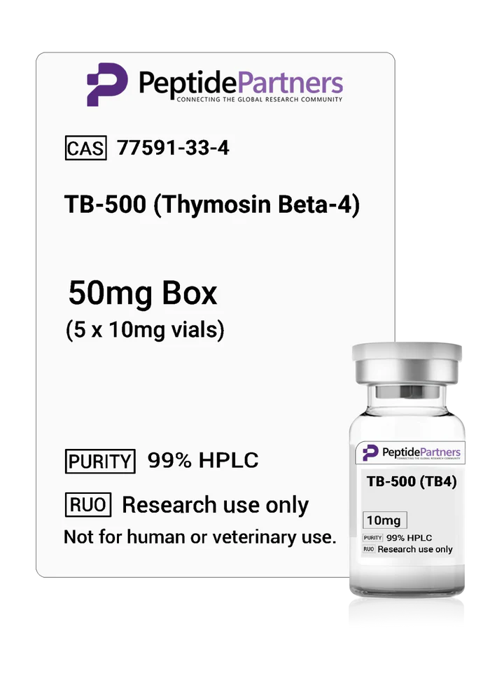
"TB-500" is the common commercial name for a synthetic research peptide modeled on thymosin beta-4. Depending on the supplier, the term may describe the full-length synthetic Tβ4 sequence or a shorter fragment built around its active actin-binding region. That ambiguity is a good reason to check the sequence, molecular weight and certificate of analysis (COA) for any lot you order. Our TB-500 (TB4) 10mg research vials are listed with the TB4 designation so researchers can match the material to their protocol.

How Thymosin Beta-4 Works: Actin Binding and Cell Migration

The best-characterized function of thymosin beta-4 is its role as a G-actin sequestering protein. Actin is the building block of the cytoskeleton, and cells constantly assemble and disassemble actin filaments as they move, divide and change shape. By binding free (monomeric) actin, Tβ4 helps regulate the pool of actin available for filament assembly.

A short central sequence of the protein, often written as LKKTETQ, is associated with this actin-binding activity. Because cell migration depends heavily on actin dynamics, researchers have used Tβ4 and related peptides to study how cells such as keratinocytes, endothelial cells and fibroblasts move into damaged tissue in experimental models. Research suggests Tβ4 may also influence signaling pathways involved in angiogenesis (new blood vessel formation) and inflammatory responses.

What Preclinical Research on TB-500 Has Examined

Most published work involves thymosin beta-4 itself rather than products sold under the TB-500 name, so findings should not be assumed to transfer automatically. Areas that have appeared in the peer-reviewed literature include:

  • Dermal wound models: rodent studies have examined Tβ4's effects on re-epithelialization, collagen deposition and angiogenesis in experimental skin wounds.
  • Cardiac research: a widely cited 2004 study in Nature reported that Tβ4 promoted cardiomyocyte migration and survival in a mouse model of cardiac injury, linked to activation of integrin-linked kinase (ILK).
  • Corneal and ocular models: Tβ4 has been studied in corneal epithelial wound models, and ophthalmic formulations of Tβ4 have been evaluated in early-phase clinical trials.
  • Inflammation signaling: in vitro and animal work has explored Tβ4's relationship with inflammatory cytokine signaling.
These are research findings in specific models. They do not establish safety or efficacy in humans, and TB-500 is not an approved drug.

TB-500 vs BPC-157 in the Research Literature

TB-500 is frequently mentioned alongside BPC-157, but the two compounds are structurally and mechanistically unrelated:

  • Origin: Tβ4 is an endogenous 43-residue protein; BPC-157 is a synthetic 15-amino-acid peptide derived from a sequence found in gastric juice protein.
  • Proposed mechanism: Tβ4 research centers on actin regulation and cell migration; BPC-157 research has focused on pathways such as nitric oxide signaling and growth factor expression in animal models.
  • Research models: Tβ4 literature leans toward dermal, cardiac and ocular models, while BPC-157 literature is heavily weighted toward gastrointestinal, tendon and ligament models in rodents.
Because their proposed mechanisms differ, some laboratories study them side by side or together. For that work we carry standalone BPC-157 10mg vials, a pre-combined BPC-157/TB-500 combo vial, and the GLOW blend with BPC-157, TB-500 and GHK-Cu.

Lab Storage and Reconstitution of Lyophilized TB-500

TB-500 is supplied as a lyophilized (freeze-dried) powder, which is considerably more stable than a peptide in solution. General laboratory handling practices for lyophilized peptides include:

  • Before reconstitution: store vials sealed, protected from light, in a freezer (around -20°C) for long-term storage or refrigerated for shorter periods.
  • Equilibrate first: let the vial reach room temperature before opening to limit moisture condensation on the powder.
  • Choose an appropriate diluent: many labs use sterile water or bacteriostatic water, which contains 0.9% benzyl alcohol to inhibit microbial growth in multi-use vials. Our Reconstitution Solution (BAC water) is available in 10mL vials.
  • Add diluent gently: run it down the inside wall of the vial and swirl rather than shake, since vigorous agitation can damage peptides.
  • After reconstitution: keep solutions refrigerated (2–8°C), label them with date and concentration, and avoid repeated freeze-thaw cycles; aliquoting is a common approach.
Always follow your institution's protocols and the guidance on the product's documentation.

TB-500 Purity, COAs and Third-Party Testing

Peptide purity directly affects the reliability of experimental results. Truncated sequences, synthesis by-products or incorrect mass can all confound data. When evaluating a TB-500 supplier, look for:

  • HPLC purity data for the specific lot, not a generic sample report
  • Mass spectrometry confirming the expected molecular weight
  • Testing performed by an independent laboratory rather than only in-house
  • Clear labeling of vial contents (for example, 10mg per vial)
You can review our independent third-party certifications to see how our peptides are tested, and our shipping information for handling and delivery details.

TB-500 Peptide FAQ

Is TB-500 the same as thymosin beta-4?
Not always. Thymosin beta-4 is the naturally occurring 43-amino-acid protein. TB-500 is a commercial name for a synthetic research peptide based on Tβ4, which may be the full sequence or a fragment depending on the supplier. Check the COA and listed sequence for the lot you purchase.

What does TB4 mean on a product label?
TB4 is shorthand for thymosin beta-4. A label such as "TB-500 (TB4)" indicates the product is a thymosin beta-4 research peptide.

How long is lyophilized TB-500 stable?
Lyophilized peptides are generally stable for extended periods when kept sealed, dry, cold and away from light. Once reconstituted, stability is much shorter, so solutions are typically refrigerated and used within the timeframe specified by the lab's own protocols.

Why do researchers compare TB-500 with BPC-157?
Both are frequently studied in tissue-repair models, but they have different structures and proposed mechanisms, which makes side-by-side comparison useful for researchers designing preclinical studies.

Order Third-Party Tested TB-500 for Your Lab

Whether your work focuses on actin dynamics, cell migration or comparative peptide studies, starting with verified, well-documented material is essential. Peptide Partners supplies TB-500 (TB4) in 10mg lyophilized vials backed by independent testing. If your lab also works with growth hormone secretagogues, see our Sermorelin vs Tesamorelin research comparison guide.

Shop TB-500 (TB4) 10mg Vials

All products are sold strictly for laboratory research use only. They are not intended for human or animal consumption, or for diagnostic or therapeutic use. Nothing in this article is medical advice.

Posted on — Leave a comment

Sermorelin vs Tesamorelin: Research Comparison Guide

Sermorelin vs tesamorelin is one of the most common comparisons researchers make when designing studies on the growth hormone–releasing hormone (GHRH) axis. Both peptides act on the same receptor, yet they differ in length, chemical stability and the body of published research behind them. This guide breaks down those differences so you can choose the right compound for your laboratory work.

Below we cover structure, mechanism, half-life, the key published studies, lab storage considerations and how to evaluate purity before you buy.

Sermorelin vs Tesamorelin at a Glance

Both compounds are synthetic GHRH analogs. Here is a quick side-by-side summary:

  • Sermorelin (GRF 1-29 NH2): a 29-amino-acid peptide corresponding to the N-terminal fragment of native human GHRH (1-44), the shortest sequence known to retain full biological activity at the GHRH receptor. Molecular weight roughly 3,358 Da.
  • Tesamorelin: the full 44-amino-acid human GHRH sequence with a trans-3-hexenoyl group attached to the N-terminal tyrosine. Molecular weight roughly 5,136 Da.
  • Shared target: the GHRH receptor (GHRH-R) on anterior pituitary somatotroph cells.
  • Main difference: tesamorelin's N-terminal modification was designed to resist enzymatic degradation, giving it greater stability than unmodified GHRH fragments.

You can review specifications for each compound on our Sermorelin 10mg research vials and Tesamorelin 10mg research vials product pages.

How GHRH Analogs Work: Shared Mechanism

Native GHRH is released by the hypothalamus and binds GHRH-R, a G protein–coupled receptor. Receptor activation raises intracellular cAMP in somatotrophs, which stimulates both synthesis and pulsatile release of growth hormone (GH). Downstream, GH drives hepatic production of insulin-like growth factor 1 (IGF-1).

A feature researchers often highlight is that GHRH analogs work upstream of GH itself. Because release still depends on pituitary function and remains subject to negative feedback from somatostatin and IGF-1, studies using GHRH analogs tend to preserve the physiological pulsatile pattern of GH secretion. That makes both sermorelin and tesamorelin useful tools for studying endogenous GH regulation, rather than models of direct GH exposure.

Structural Differences and Stability

The biggest practical distinction between the two peptides is stability. Native GHRH and short fragments like sermorelin are rapidly cleaved by dipeptidyl peptidase-4 (DPP-4) between positions 2 and 3, producing an inactive metabolite. Published pharmacology describes sermorelin's plasma half-life as on the order of minutes.

Tesamorelin was engineered specifically to address this. The trans-3-hexenoyl group on its N-terminus protects that cleavage site, and research describes a longer half-life and more sustained GHRH-R activation than unmodified GHRH. For researchers, this means:

  • Sermorelin suits experiments focused on short, discrete stimulation of the GHRH receptor.
  • Tesamorelin suits study designs where a more stable, longer-acting full-length analog is needed.
  • Both differ from tetrasubstituted analogs such as CJC-1295 no DAC (Modified GRF 1-29), which uses four amino acid substitutions to improve the stability of the 1-29 fragment.

Published Research on Sermorelin

Sermorelin has a long research history. It was developed in the 1980s and was previously marketed in the United States as a diagnostic agent for assessing pituitary GH reserve; the manufacturer later discontinued it for commercial reasons. Published literature has examined sermorelin in studies of GH secretory dynamics, pituitary responsiveness testing, and age-related changes in the GH/IGF-1 axis. Because its sequence matches the active core of native GHRH, it is frequently used as a reference compound when characterizing newer GHRH analogs.

Published Research on Tesamorelin

Tesamorelin has a more recent and extensive clinical research record. Large randomized controlled trials examined it in adults with HIV-associated lipodystrophy, measuring visceral adipose tissue by CT imaging, and these studies led to its 2010 FDA approval for that specific indication. Subsequent research has investigated its effects on liver fat in HIV-associated nonalcoholic fatty liver disease, as well as markers of body composition and IGF-1 response.

For investigators, this body of work provides well-characterized pharmacodynamic data, which can be valuable when designing preclinical models of the GHRH axis and visceral fat metabolism.

Choosing Between Sermorelin and Tesamorelin for Your Lab

When deciding which GHRH analog fits your protocol, consider:

  • Study duration: short-acting stimulation studies often favor sermorelin; longer-exposure designs may favor tesamorelin's improved stability.
  • Reference literature: tesamorelin has more recent large-scale clinical data; sermorelin has decades of foundational pharmacology.
  • Sequence requirements: if your work requires the full 1-44 GHRH sequence, tesamorelin is the closer match.
  • Comparative designs: some labs run both side by side, or alongside a ghrelin-receptor agonist such as those in our CJC-1295 no DAC/Ipamorelin combo vial, to compare GHRH-R and GHS-R1a pathways.

Whichever compound you choose, purity matters. Impurities, truncated sequences or incorrect peptide content can skew receptor-activation data. Look for HPLC and mass spectrometry results, and review our independent third-party certifications before ordering. Both peptides ship as lyophilized powder; for lab handling they are typically stored frozen and protected from light, then reconstituted with a sterile diluent such as our Reconstitution Solution (BAC water) and kept refrigerated.

FAQ: Sermorelin vs Tesamorelin

Is tesamorelin just a longer version of sermorelin?
Not exactly. Sermorelin is the 1-29 fragment of GHRH, while tesamorelin is the full 1-44 sequence plus an N-terminal trans-3-hexenoyl modification that improves resistance to enzymatic breakdown.

Do sermorelin and tesamorelin act on the same receptor?
Yes. Both are GHRH receptor agonists. They differ mainly in structure, stability and the published research associated with each, not in their primary target.

How is sermorelin different from CJC-1295?
CJC-1295 no DAC (Modified GRF 1-29) is based on the same 1-29 fragment as sermorelin but includes four amino acid substitutions intended to increase stability against degradation.

How should lyophilized GHRH peptides be stored in the lab?
Lyophilized peptides are generally stored frozen, sealed and away from light and moisture. After reconstitution, solutions are typically refrigerated and used within the timeframe your lab's protocol specifies. See our shipping information for how orders are packed and delivered.

Source Third-Party Tested GHRH Peptides

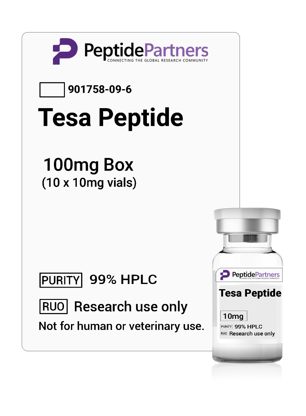
Comparing sermorelin vs tesamorelin comes down to your study design: the shorter, classic 1-29 fragment or the stabilized full-length analog. Peptide Partners supplies both in 10mg research vials, backed by independent third-party testing. Browse the product page to review specifications and documentation for your next study.

Shop Tesamorelin 10mg Research Vials

All products are sold strictly for laboratory research use only. They are not intended for human or animal consumption, or for diagnostic or therapeutic use. Nothing in this article is medical advice.